Anatomy Hip Muscles Diagram / Hip Anatomy And Function. Diagram only (coloring book quiz) learn with flashcards, games and more — for free. Use the mouse scroll wheel to move the images up and down alternatively use the tiny arrows (>>) on both side of the image to move the images. It allows us to walk, run, and jump. Comprehensive information about hip joint anatomy including muscles, tendons, ligaments, bones, bursae, skeletal structure and joint capsules. Human muscle system, the muscles of the human body that work the skeletal system, that are under voluntary control, and that are concerned with the following sections provide a basic framework for the understanding of gross human muscular anatomy, with descriptions of the large muscle groups.
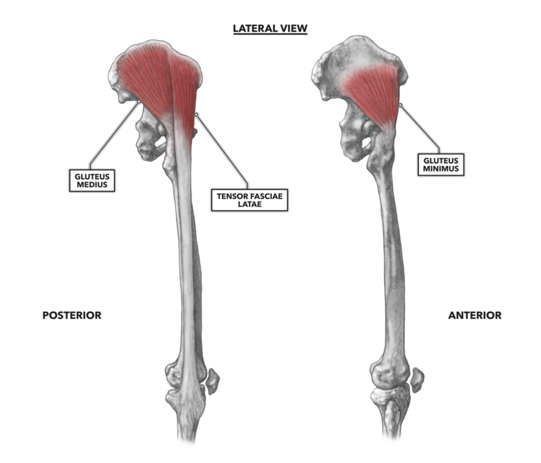
Diagram represnting the posterier view of the hamstring muscle group. In human anatomy, the muscles of the hip joint are those muscles that cause movement in the hip. 1 hip anatomy, function and common problems. Microscopic anatomy of skeletal muscle. Muscles of hip and thigh lateral view.
Groin Muscle Anatomy Diagram Groin Muscle Anatomy Diagram Muscles On Inner Thigh Best Inner Thigh Stre Body Muscle Anatomy Muscle Anatomy Hip Muscles Anatomy from i.pinimg.com The hip muscles work together to carry out 4 different types of movement: The hip's unique anatomy enables it to be both extremely strong and amazingly flexible, so it can bear weight and allow for a wide range of movement. Most modern anatomists define 17 of these muscles, although some additional muscles may sometimes be considered. Attached to the bones of the skeletal system are about 700 named muscles that make up roughly half of a person's body weight. Muscles that act on the lower limb cause movement at the hip, knee and foot joints. This tutorial will teach you all about the six hip adductor muscles. Related posts of muscles of the lower back and hip diagram muscle anatomy posterior. Bursae of the lower limb:
It allows us to walk, run, and jump.
Knowing the anatomy of your hip can help you understand the source of any hip pain. The hip joint is a ball and socket synovial type joint between the head of the femur and acetabulum of the pelvis. This mri hip joint axial cross sectional anatomy tool is absolutely free to use. Use the mouse scroll wheel to move the images up and down alternatively use the tiny arrows (>>) on both side of the image to move the images. The muscles of the pelvis, hip and buttock anatomical chart shows how each muscle in this area of the body works with the others, and the various minor systems within the major ones. Diagram represnting the posterier view of the hamstring muscle group. The different bursae of the hip region (trochanteric, ischial and. Posted on january 20, 2015 by admin. Practically all muscles in this group have the. Related posts of muscles of the lower back and hip diagram muscle anatomy posterior. The quadricep group of muscles is the muscles found on the anterior portion of the thigh and they are the rectus femoris, the lateralis, the vastus medialis, and the. You can click the links in the image, or the links below the image to find out more information on any muscle group. The anatomy of the fascia lata and iliotibial tract.
Use the mouse scroll wheel to move the images up and down alternatively use the tiny arrows (>>) on both side of the image to move the images. Diagram only (coloring book quiz) learn with flashcards, games and more — for free. Muscles that act on the lower limb cause movement at the hip, knee and foot joints. Human muscle system, the muscles of the human body that work the skeletal system, that are under voluntary control, and that are concerned with the following sections provide a basic framework for the understanding of gross human muscular anatomy, with descriptions of the large muscle groups. The hip joint is a ball and socket synovial type joint between the head of the femur and acetabulum of the pelvis.
Crossfit Hip Musculature Part 3 Lateral Muscles from www.crossfit.com The hip has several ligaments connecting the femur to the pelvis and tendons connecting the bones to many surrounding muscles. Thats what the m stands for. The hip's unique anatomy enables it to be both extremely strong and amazingly flexible, so it can bear weight and allow for a wide range of movement. The anatomy of the fascia lata and iliotibial tract. These two muscles are often associated as one muscle since one is generally nearly useless without the other. Muscles that act on the lower limb cause movement at the hip, knee and foot joints. The anterior muscles of the hip allow for rotational movements and flexion of the hip as well as flexion of the vertebral column, but only when they apply their contraction during cohesive unison. The hip joint ( coxa in latin) is the articulation connecting the pelvis and the femur.
Use the mouse scroll wheel to move the images up and down alternatively use the tiny arrows (>>) on both side of the image to move the images.
Use the mouse scroll wheel to move the images up and down alternatively use the tiny arrows (>>) on both side of the image to move the images. Thats what the m stands for. The hip has several ligaments connecting the femur to the pelvis and tendons connecting the bones to many surrounding muscles. The quadricep group of muscles is the muscles found on the anterior portion of the thigh and they are the rectus femoris, the lateralis, the vastus medialis, and the. We all have a layer of fatty tissue under our skin, and this softens the look of the underlying muscles. And you'll be in a better position to help your doctor pinpoint the cause. Microscopic anatomy of skeletal muscle. Human anatomy hip muscles anatomy anatomy study. Find the best weight lifting exercises that target each muscle or groups of muscles. Knowing the anatomy of your hip can help you understand the source of any hip pain. In human anatomy, the muscles of the hip joint are those muscles that cause movement in the hip. Learn more about the anatomy of the hip using these hip diagrams that will show you the detailed structure of your hip! The hip's unique anatomy enables it to be both extremely strong and amazingly flexible, so it can bear weight and allow for a wide range of movement.
Diagram only (coloring book quiz) learn with flashcards, games and more — for free. It bears our body's weight and the force of the strong muscles of the hip and leg. Included within the chart are gorgeous illustrations of the pelvic diaphragm, sphincter muscles, gluteus maximus. The hip's unique anatomy enables it to be both extremely strong and amazingly flexible, so it can bear weight and allow for a wide range of movement. The anterior muscles of the hip allow for rotational movements and flexion of the hip as well as flexion of the vertebral column, but only when they apply their contraction during cohesive unison.
Understand Hip Anatomy Muscles For Yoga Jason Crandell Yoga from www.jasonyoga.com It allows us to walk, run, and jump. Find the best weight lifting exercises that target each muscle or groups of muscles. Attached to the bones of the skeletal system are about 700 named muscles that make up roughly half of a person's body weight. Thats what the m stands for. Muscle and tendon anatomy of the hip (adductors, gluteal muscles (or buttocks), hamstring muscles, femoral muscle quadrices). #back muscles diagram #body muscles diagram labeled #diagram of hip muscles and ligaments #hip anatomy diagram #hip muscles pain #thigh muscles diagram. Muscles of hip and thigh lateral view. Muscle movements, types, and names.
Find the best weight lifting exercises that target each muscle or groups of muscles.
Common hip and back pain causes include injury to muscles from overuse disc injurydegeneration or spinal stenosis. The six hip adductor muscles are all located in the adductor or medial compartment of the thigh and all mainly adduct the thigh at the hip joint. We all have a layer of fatty tissue under our skin, and this softens the look of the underlying muscles. The anatomy of the fascia lata and iliotibial tract. Related posts of muscles of the lower back and hip diagram muscle anatomy posterior. Muscle and tendon anatomy of the hip (adductors, gluteal muscles (or buttocks), hamstring muscles, femoral muscle quadrices). The hip joint is one of the most important joints in the human body. It takes great force to seriously damage the hip because of the strong, large muscles of the thighs that support and move the hip. The hip joint ( coxa in latin) is the articulation connecting the pelvis and the femur. It allows us to walk, run, and jump. Diagram represnting the posterier view of the hamstring muscle group. The hip muscles work together to carry out 4 different types of movement: Discover the muscle anatomy of every muscle group in the human body.
Rectus femoris, named for its muscle fascicle orientation due to its muscular orientation, it causes flexion and lateral rotation at the hip and knee flexion hip muscles diagram. It takes great force to seriously damage the hip because of the strong, large muscles of the thighs that support and move the hip.
Share :
Post a Comment
for "Anatomy Hip Muscles Diagram / Hip Anatomy And Function"


Post a Comment for "Anatomy Hip Muscles Diagram / Hip Anatomy And Function"